

What Modifies And Packages Proteins
5.vi: Jail cell Organelles
- Page ID
- 16744
Figure \(\PageIndex{1}\) represents an important construction in living cells. It is a component of a ribosome, the prison cell structure where proteins are synthesized. Large ribosomal subunit (50S) of Haloarcula marismortui, facing the 30S subunit. The ribosomal proteins are shown in blue, the rRNA in ochre (a shade of brown and yellow), the active site in red. All living cells contain ribosomes, whether they are prokaryotic or eukaryotic cells. However, but eukaryotic cells too contain a nucleus and several other types of organelles.

An organelle is a construction within the cytoplasm of a eukaryotic prison cell that is enclosed within a membrane and performs a specific task. Organelles are involved in many vital cell functions. Organelles in brute cells include the nucleus, mitochondria, endoplasmic reticulum, Golgi apparatus, vesicles, and vacuoles. Ribosomes are non enclosed inside a membrane simply are still unremarkably referred to as organelles in eukaryotic cells.
The Nucleus
The nucleus is the largest organelle in a eukaryotic cell and is considered to be the prison cell's command center. It contains most of the prison cell's DNA, which makes upwards chromosomes and is encoded with the genetic instructions for making proteins. The function of the nucleus is to regulate factor expression, including controlling which proteins the cell makes. In add-on to Dna, the nucleus contains a thick liquid called nucleoplasm that is similar in composition to the cytosol found in the cytoplasm outside the nucleus (Figure \(\PageIndex{ii}\)). Most eukaryotic cells contain but a single nucleus, simply some types of cells, such as reddish claret cells, contain no nucleus. A few other types of cells, such as muscle cells, comprise multiple nuclei.

As you tin can see from the model in Effigy \(\PageIndex{two}\), the membrane enclosing the nucleus is called the nuclear envelope. This is actually a double membrane that encloses the entire organelle and isolates its contents from the cellular cytoplasm. Tiny holes, called nuclear pores, let big molecules to pass through the nuclear envelope with the aid of special proteins. Large proteins and RNA molecules must be able to pass through the nuclear envelope and then proteins tin can be synthesized in the cytoplasm and the genetic fabric can be maintained inside the nucleus. The nucleolus shown in the model beneath is mainly involved in the associates of ribosomes. After being produced in the nucleolus, ribosomes are exported to the cytoplasm where they are involved in the synthesis of proteins.
Mitochondria
The mitochondrion (plural, mitochondria) is an organelle that makes energy available to the prison cell (Figure \(\PageIndex{three}\)). This is why mitochondria are sometimes referred to as the power plants of the cell. They use energy from organic compounds such as glucose to make molecules of ATP (adenosine triphosphate), an free energy-carrying molecule that is used almost universally inside cells for free energy.
Scientists think that mitochondria were one time complimentary-living organisms because they contain their own Deoxyribonucleic acid. They theorize that ancient prokaryotes infected (or were engulfed by) larger prokaryotic cells, and the 2 organisms evolved a symbiotic relationship that benefited both of them. The larger cells provided the smaller prokaryotes with a place to live. In return, the larger cells got actress energy from the smaller prokaryotes. Eventually, the smaller prokaryotes became permanent guests of the larger cells, every bit organelles inside them. This theory is called the endosymbiotic theory, and it is widely accepted by biologists today
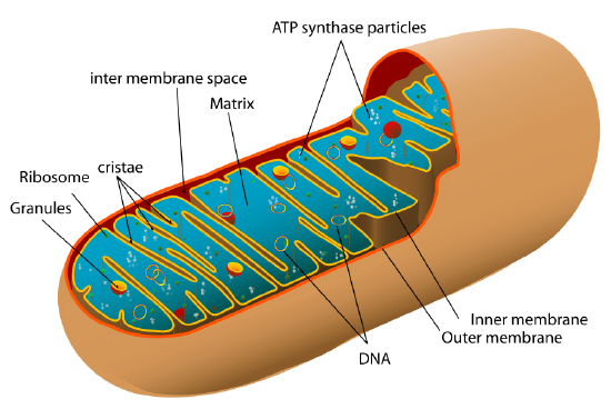
Mitochondrial Compartments
The double membrane nature of the mitochondria results in five distinct compartments, each with an of import function in cellular respiration. These compartments are:
- the outer mitochondrial membrane,
- the intermembrane space (the infinite between the outer and inner membranes),
- the inner mitochondrial membrane,
- the cristae (formed by infoldings of the inner membrane), and
- the matrix (space inside the inner membrane).
Endoplasmic Reticulum
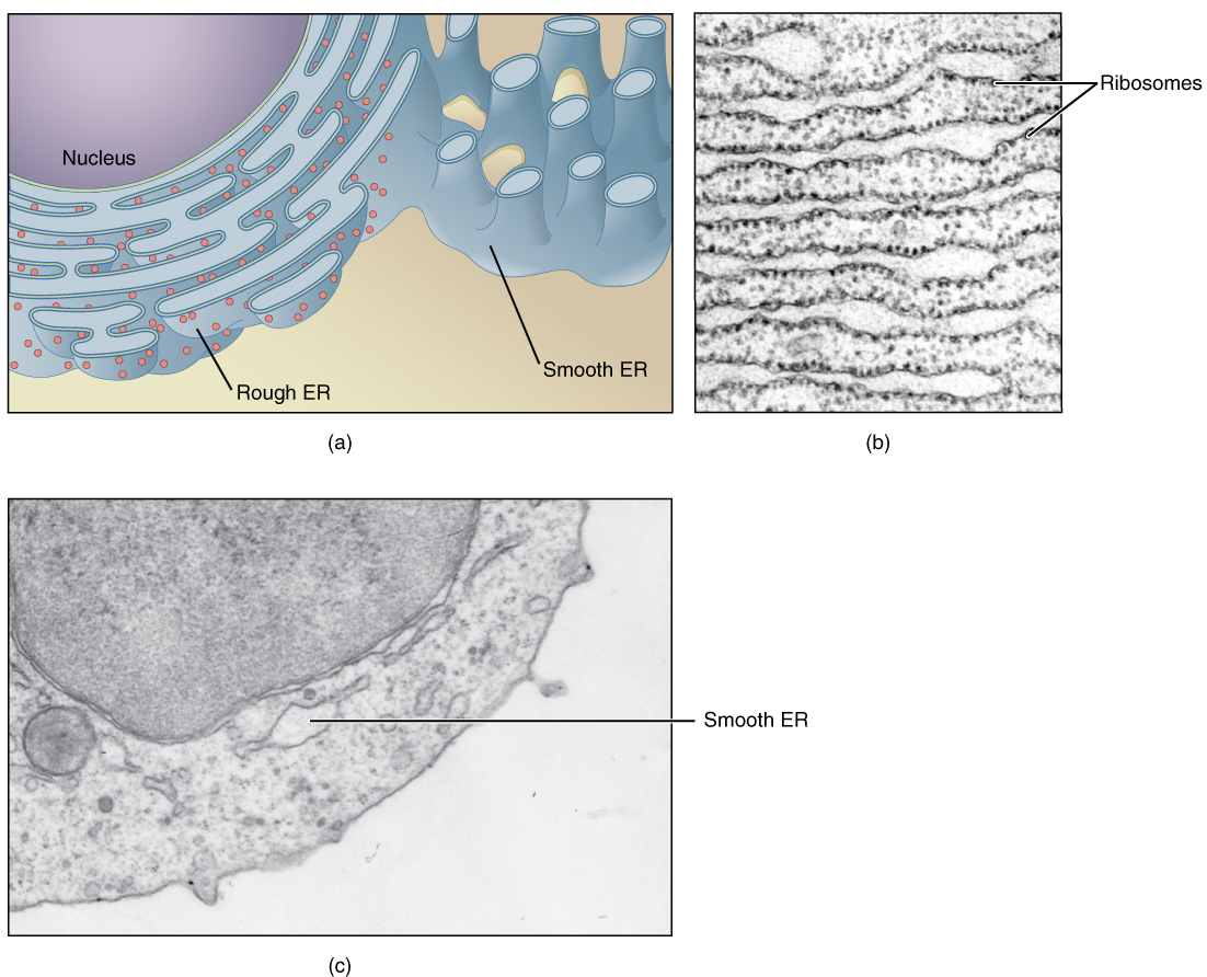
The endoplasmic reticulum (ER) (plural, reticuli) is a network of phospholipid membranes that form hollow tubes, flattened sheets, and round sacs. These flattened, hollow folds and sacs are called cisternae. The ER has two major functions:
- Transport: Molecules, such as proteins, can move from place to place inside the ER, much similar on an intracellular highway.
- Synthesis: Ribosomes that are attached to the ER, like to unattached ribosomes, make proteins. Lipids are also produced in the ER.
There are two types of endoplasmic reticulum, rough endoplasmic reticulum (RER) and smooth endoplasmic reticulum (SER):
- Rough endoplasmic reticulum is studded with ribosomes, which gives it a "crude" advent. These ribosomes brand proteins that are then transported from the ER in small sacs called transport vesicles. The transport vesicles compression off the ends of the ER. The rough endoplasmic reticulum works with the Golgi apparatus to move new proteins to their proper destinations in the cell. The membrane of the RER is continuous with the outer layer of the nuclear envelope.
- Smoothen endoplasmic reticulum does not have any ribosomes attached to it, and so it has a polish appearance. SER has many different functions, some of which include lipid synthesis, calcium ion storage, and drug detoxification. The shine endoplasmic reticulum is plant in both animal and found cells and information technology serves dissimilar functions in each. The SER is made up of tubules and vesicles that branch out to class a network. In some cells, there are dilated areas like the sacs of RER. Smooth endoplasmic reticulum and RER form an interconnected network.
Golgi Apparatus
The Golgi appliance (Figure \(\PageIndex{five}\)) is a big organelle that processes proteins and prepares them for use both inside and outside the cell. Information technology was identified in 1898 past the Italian dr. Camillo Golgi. The Golgi appliance modifies, sorts, and packages different substances for secretion out of the jail cell, or for use within the jail cell. The Golgi apparatus is institute close to the nucleus of the cell where it modifies proteins that have been delivered in transport vesicles from the Rough Endoplasmic Reticulum. It is likewise involved in the send of lipids around the cell. Pieces of the Golgi membrane pinch off to form vesicles that transport molecules effectually the prison cell. The Golgi apparatus tin exist idea of equally similar to a postal service function; it packages and labels "items" then sends them to different parts of the jail cell. The Golgi apparatus tends to exist larger and more numerous in cells that synthesize and secrete large quantities of materials; for instance, the plasma B cells and the antibiotic-secreting cells of the immune system take prominent Golgi complexes.
The Golgi apparatus manipulates products from the Crude Endoplasmic Reticulum (ER) and likewise produces new organelles called lysosomes. Proteins and other products of the ER are sent to the Golgi apparatus, which organizes, modifies, packages, and tags them. Some of these products are transported to other areas of the cell and some are exported from the prison cell through exocytosis. Enzymatic proteins are packaged as new lysosomes.
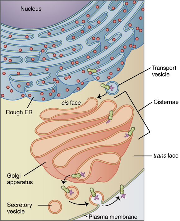
The stack of cisternae has four functional regions: the cis-Golgi network, medial-Golgi, endo-Golgi, and trans-Golgi network. Vesicles from the ER fuse with the network and subsequently progress through the stack from the cis- to the trans-Golgi network, where they are packaged and sent to their destination. Each cisterna includes special Golgi enzymes which modify or help to modify proteins that travel through information technology. Proteins may be modified past the addition of a sugar group (glycosylation) or phosphate group (phosphorylation). These modifications may form a signal sequence on the protein, which determines the final destination of the protein. For case, the addition of mannose-6-phosphate signals the protein for lysosomes.
Vesicles and Vacuoles
Both vesicles and vacuoles are sac-like organelles that store and transport materials in the cell. Vesicles are much smaller than vacuoles and have a multifariousness of functions. The vesicles that compression off from the membranes of the ER and Golgi apparatus shop and ship protein and lipid molecules. You tin can see an example of this blazon of transport vesicle in the figure above. Some vesicles are used as chambers for biochemical reactions. Other vesicles include:
- Lysosomes, which use enzymes to break down strange matter and dead cells.
- Peroxisomes, which use oxygen to interruption downwards poisons.
- Transport vesicles, transport contents between organelle also equally between prison cell exterior and interior.
Centrioles
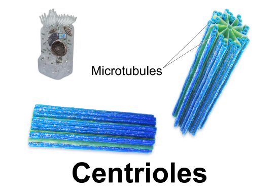
Centrioles are organelles involved in cell partition. The function of centrioles is to help organize the chromosomes before cell division occurs and then that each daughter cell has the right number of chromosomes later on the cell divides. Centrioles are found only in brute cells and are located nearly the nucleus. Each centriole is made mainly of a poly peptide named tubulin. The centriole is cylindrical in shape and consists of many microtubules, as shown in the model pictured beneath.
Ribosomes
Ribosomes are small structures where proteins are made. Although they are non enclosed within a membrane, they are frequently considered organelles. Each ribosome is formed of ii subunits, like the one pictured at the top of this section. Both subunits consist of proteins and RNA. RNA from the nucleus carries the genetic code, copied from DNA, which remains in the nucleus. At the ribosome, the genetic code in RNA is used to assemble and join together amino acids to make proteins. Ribosomes can be institute alone or in groups within the cytoplasm as well as on the RER.
Review
- Define organelle.
- Draw the construction and part of the nucleus.
- Explicate how the nucleus, ribosomes, rough endoplasmic reticulum, and Golgi apparatus work together to make and transport proteins.
- Why are mitochondria referred to as the power plants of the cell?
- What roles are played by vesicles and vacuoles?
- Why do all cells demand ribosomes, even prokaryotic cells that lack a nucleus and other cell organelles?
- Explain endosymbiotic theory as information technology relates to mitochondria. What is ane piece of evidence that supports this theory?
- Lysosomes and peroxisomes are types of:
- A. Organelles
- B. Vesicles
- C. Vacuoles
- D. Both A and B
- Which of the following organelles fits best with each description of function? Choose merely one organelle for each answer: Golgi apparatus, centrioles, nucleolus, nucleus, rough endoplasmic reticulum
- a. Contains the genetic instructions for the production of proteins
- b. Organizes chromosomes before cell sectionalisation
- c. Provides a framework for ribosomes
- d. Packages and labels proteins
- e. Assembles ribosomes
- True or False. All eukaryotic cells have a nucleus.
- True or False. The outer surface of the nucleus of a eukaryotic cell is not completely solid.
Explore More

What Modifies And Packages Proteins,
Source: https://bio.libretexts.org/Bookshelves/Human_Biology/Book%3A_Human_Biology_(Wakim_and_Grewal)/05%3A_Cells/5.06%3A_Cell_Organelles
Posted by: porternoust1988.blogspot.com
0 Response to "What Modifies And Packages Proteins"
Post a Comment